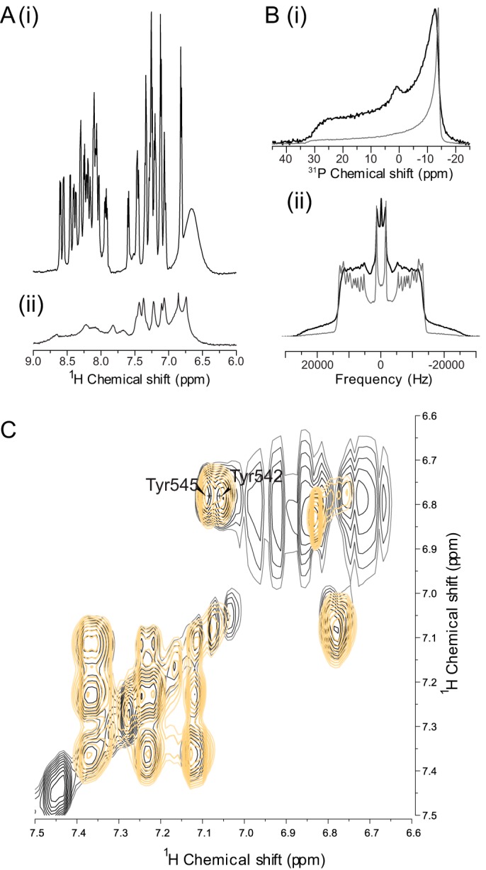
FIGURE 2.

Tyrosine residues within the S4S5 linker of Kv11.1 are oriented toward the lipid membrane. A, S4S5 peptide (Leu-532–Phe-551) interacts with the lipid membrane as the amide and aromatic resonances in the 1H NMR spectrum of the WT peptide in DMPC/DPC-d38 are very broad (panel ii) compared with S4S5 peptide in 10% D2O (panel i). B, pure 31P (panel i) and 2H (panel ii) solid-state NMR spectra of DMPC-d54 MLV model membranes (gray) and with S4S5 peptide (black) showing the S4S5 peptide interacts with the lipid membrane. C, 1H TOCSY spectra of the S4S5 peptide solubilized with DMPC-d54/DPC-d38 in 10% D2O (black) and with 0.5 mm Gd3+ (orange) showing the tyrosine residues (Tyr-542 and Tyr-545) are protected from soluble paramagnetics by the lipids as the aromatic 1H signals for the Tyr-542 and Tyr-545 remain.
