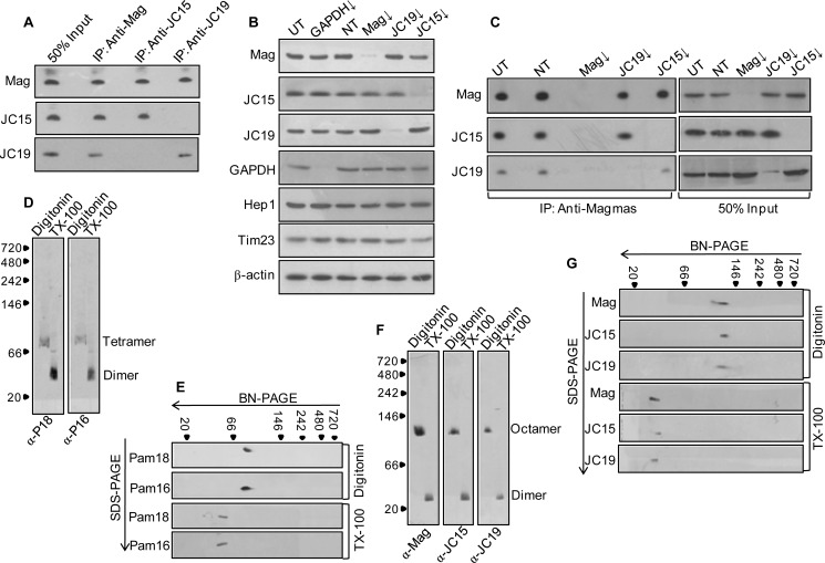
FIGURE 1.
Assembly of import motor associated J-protein subcomplexes at the translocation channel. A, Triton X-100-lysed mitochondria were subjected to immunoprecipitation using anti-Magmas, anti-JC19, or anti-JC15 antibodies. The interacting proteins were probed by immunoblotting using specific antibodies. 50% of soluble material after lysis was used as the loading control (50% Input). B, HEK293T cells were seeded in Opti-MEM media and transfected with 5 μm siRNA pool against Magmas (Mag), JC19, and JC15 using Lipofectamine 2000. After 48 h, mitochondria were isolated and probed for expression of individual proteins. siGAPDH was used as a positive control, Hep1 was the mitochondrial loading control, β-actin was the initial cell concentration control, Tim23 was a control for translocase integrity. C, the assembly of J-protein subcomplex upon knockdown of individual protein components was assayed by subjecting the isolated mitochondria to co-immunoprecipitation analysis using anti-Magmas antibodies. The immunoprecipitates were separated on SDS-PAGE and probed with antibodies specific to Magmas, JC15, and JC19. 50% of the soluble material after lysis was used as a loading control (50% Input). D and F, 1% Triton X-100 or 1% digitonin-lysed human mitochondria was resolved on blue native gel and immunoblotted with Magmas-, JC19-, or JC15-specific antibodies. To decipher the oligomeric state, the relative migration of the bands was mapped onto the molecular weight standard. To correlate, equivalently treated yeast mitochondria (D) was subjected to blue native PAGE and immunoblotted with Pam16 or Pam18 antibodies. E and G, analysis of J-protein oligomerization through two-dimensional immunoblot of Triton X-100 (TX-100) and digitonin-lysed yeast (E) and human (G) mitochondria. Mitochondrial lysates were resolved on blue native (BN-PAGE) PAGE followed by second dimension separation on SDS-PAGE. Individual components of the oligomers were determined by staining the blot with respective antibodies. UT, untransfected control; NT, transfected with non-targeting dsiRNA as internal control; ↓, mRNA down-regulated by dsiRNA).

