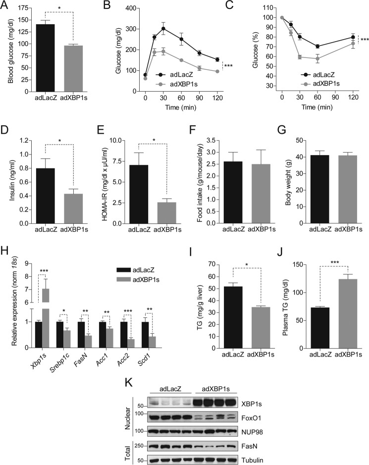
FIGURE 1.
XBP1s reduces hepatic triglyceride content and lipogenic gene expression. C57BL6/J mice (3 weeks old) were fed an HFD (45% kcal) for 12 weeks and injected with adLacZ, adXBP1s, or ΔDBD-XBP1s (8*10E7 pfu/g) through the tail vein. A, blood glucose levels were assessed after 6 h of fasting on post-adenovirus injection day 9. B, a GTT with 1 g/kg glucose (i.p. injection) was performed on day 5 post-adenovirus injection. C, an ITT was performed on day 7 post-adenovirus injection by intraperitoneal insulin (0.75 IU/kg) injection. D, plasma insulin levels after 6-h fasting on day 9 post-adenovirus injection. E, homeostatic model assessment for insulin resistance as calculated from 6 h-fasted glucose levels and corresponding plasma insulin levels on day 9 post-adenovirus injection. F, average daily food intake (g/mouse/day) and G, body weight (g). H, relative hepatic mRNA levels of Xbp1s, Srebp1c, FasN, Acc1, Acc2, and Scd1 in livers of adLacZ and adXBP1s-injected mice. Gene expression was normalized using the 18s gene as a housekeeping gene. I and J, liver triglyceride content (I) and plasma triglyceride levels (J) were determined in 6 h-fasted mice on post-injection day 9. K, immunoblotting of XBP1s and FoxO1 in the nucleus and FasN in total homogenates of livers of LacZ and XBP1s-expressing mice. NUP98 was used as a loading control for nuclear lysates, whereas Tubulin served as a loading control for total liver lysates. Error bars are represented as mean ± S.E., and p values were determined by Student's t test or two-way analysis of variance testing with Bonferroni post-test for multiple comparison analysis (for GTT and ITT). *, p < 0.05; **, p < 0.01; ***, p < 0.001.

