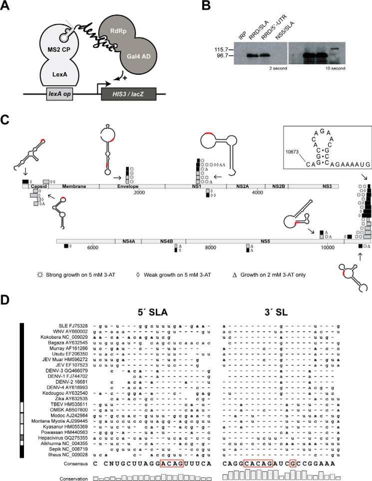
FIGURE 1.
Interaction landscape of DENV RdRp with the entire RNA genome. A, schematic diagram of Y3H in this study. B, Western blotting with polyclonal anti-NS5 antibodies (Gentex 103350) confirmed the expression of DENV-2 NS5 (120.9 kDa) and its RdRp (90.1 kDa) in Y3H with two representative RNAs, SLA and the full-length 5′-UTR. The protein IRP in pACT2 was used as a negative control. C, DENV genome is depicted as light gray rectangles and is numbered. The coding regions for viral proteins are labeled. Positive strand RNA fragments of the genome that bind the full-length NS5 and RdRp are shown above the genome as black and gray rectangles, respectively, whereas negative fragments are below. Levels of yeast growth in the presence of 3-AT are indicated by three symbols. Representative Mfold structures of RNA fragments are drawn with the CACAG motif in red. D, conservation of the ACAG loop motif is compared between the top loop regions of the 5′-SLA and the 3′-SL. Sequences are ordered according to a phylogenetic tree based on the terminal 120 nucleotides of Flaviviridae. The left vertical bar indicates the type of Flaviviridae as follows: black indicates mosquito-borne viruses; white indicates tick-borne viruses; light gray indicates viruses of no known vector; and dark gray indicates a single hepacivirus example. Consensus nucleotides are indicated once at the bottom of the figure; elsewhere, they are indicated by hyphens, with variations from the consensus indicated by the appropriate letter.

