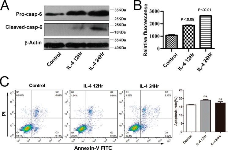
FIGURE 4.
Caspase-6 is not an apoptosis executor in alternatively M2-activated macrophages. RAW264.7 cells were stimulated with IL-4 (10 ng/ml) for 12 or 24 h, respectively. A, protein level of pro-Caspase-6 and cleaved Caspase-6 all were up-regulated in IL-4-induced macrophages; the results were detected by Western blotting with the specific antibodies. B, activity of Caspase-6 was measured using an enzymatic activity assay kit. C, apoptosis of M2 was detected by FLC. The percentages of early or late apoptosis are presented in the lower right and upper right quadrants, respectively. Columns represent the average proportions of apoptotic cells. All bars were expressed as means ±S.D. (p values of plotted data of ≤0.05 were considered statistically significant). ns indicates not significantly different.

