FIGURE 7.
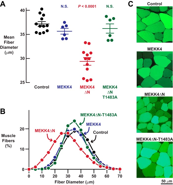
MEKK4ΔN induces skeletal muscle fiber atrophy in a Gadd45a-independent manner. A–C, mouse TA muscles were transfected with 5 μg of empty pcDNA plasmid, 5 μg of MEKK4-FLAG plasmid, 5 μg of MEKK4ΔN-FLAG plasmid, and/or 5 μg of MEKK4ΔN-T1483A-FLAG plasmid, as indicated. All muscles were co-transfected with 2.5 μg of eGFP plasmid. TA muscles were harvested for histological analysis 7 days post-transfection. A, average diameters of skeletal muscle fibers. Each data point represents the mean of >400 muscle fibers from one muscle, and horizontal bars denote average of the means ± S.E. p values were determined with a one-way ANOVA and Dunnett's multiple comparison test. N.S., not significant or p > 0.05. B, size distribution of all muscle fibers from A. C, representative fluorescence microscopy images of muscle cross-sections.

