
FIGURE 3.
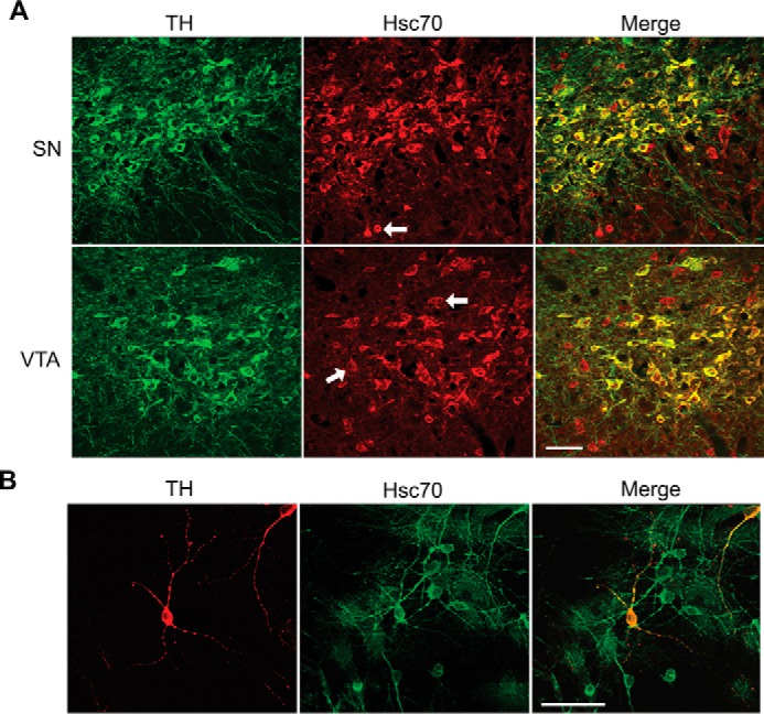
Hsc70 and TH co-localize in brain dopamine neurons. A, mouse midbrain sections of the substantia nigra (SN) and ventral tegmental area (VTA) were immunostained with polyclonal anti-TH (green, labeled with Alexa Fluor 488) and monoclonal Hsc70 (red, labeled with Alexa Fluor 555) antibodies. B, primary cell cultures from rat midbrain were immunostained with a polyclonal anti-TH (red, labeled with Alexa Fluor 555) and monoclonal Hsc70 antibodies (green, labeled with Alexa Fluor 488) antibodies. In both samples, merged images show that a majority of the TH-positive cells were dual-labeled with Hsc70 in both preparations (A and B, right panels). Only a few TH cells did not contain Hsc70. The co-localization of TH and Hsc70 extended into the process of the dopaminergic neurons. (B, right panels). Scale bars = 50 μm.