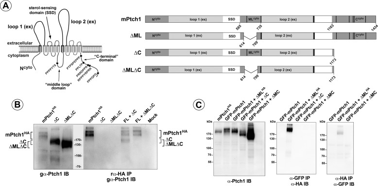
FIGURE 1.
Ptch1 structure and oligomerization. A, left panel, the predicted structure of the 12-pass transmembrane protein, Ptch1. Arrows indicate significant domains within the protein structure, and polyproline motifs are denoted in red. Right panel, the large deletion mutants of Ptch1 missing, either ΔML, ΔC, or ΔMC. Numbers refer to amino acids. B, left panel, the straight Western blot detecting HA-tagged full-length Ptch1, ΔC, and ΔMLΔC Ptch1 mutant proteins. Right panel, the ΔC mutant, but not the ΔMLΔC mutant, co-immunoprecipitates with full-length Ptch1. IB, immunoblot; IP, immunoprecipitation. C, GFP-tagged full-length Ptch1 transiently transfected with an HA-tagged ΔML mutant. Both α-GFP and reciprocal α-HA co-immunoprecipitations confirm that the ΔML deletion mutant of Ptch1 associates with full-length Ptch1. SSD, sterol sensing domain.

