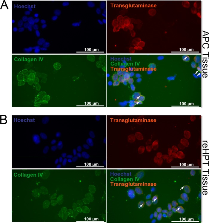
FIGURE 5.
Co-localization of extracellular TGase and collagen IV on the surface of APC and reHPT tissues. A, double staining for extracellular TGase and collagen IV, a component of the extracellular matrix and a substrate of TGase, was performed on partially digested APC tissue. B, double staining for extracellular TGase and collagen IV was performed on partially digested reHPT tissue. Hoechst 33258 (blue) was used as a nuclear stain. Arrows indicate co-localization of TGase and collagen IV on the surface of APC and reHPT cells.

