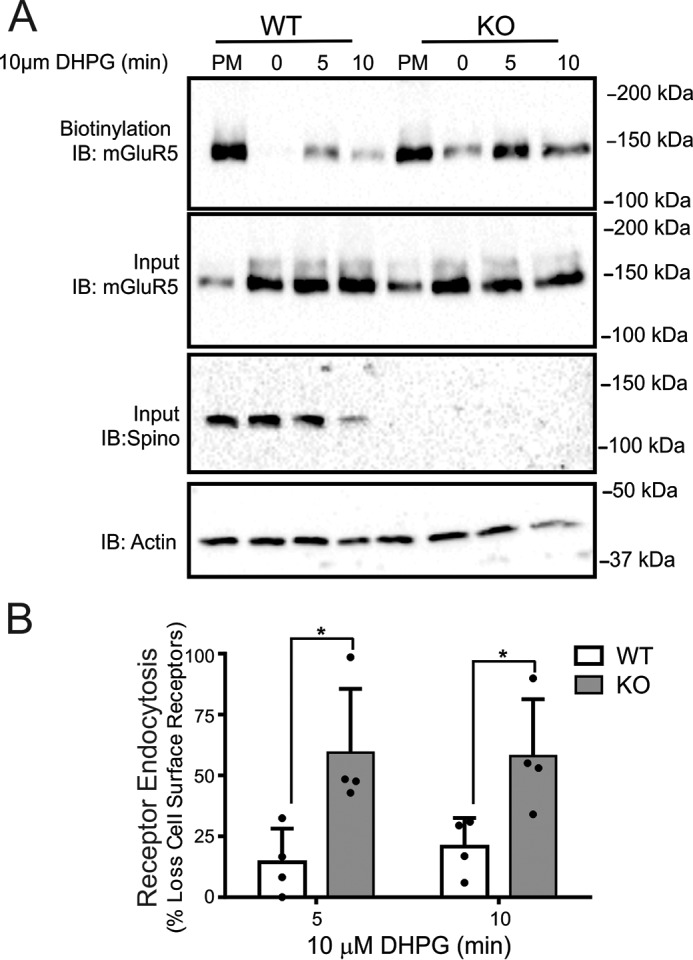
FIGURE 3.

Regulation of Group I mGluR endocytosis in cortical neurons derived from wild-type and spinophilin knock-out mice. A, the top blot shows a representative immunoblot (IB) for internalized biotin-labeled endogenous mGluR5 in primary cortical neurons (12–14 DIV) derived from E18 wild-type and spinophilin knock-out embryos in response to 10 μm DHPG treatment for 0, 5, and 10 min. The bottom blots show cell lysates (50 μg) for endogenous mGluR5 and spinophilin protein expression. B, the bar graph shows the densitometric analysis of internalized biotin-labeled mGluR5 protein normalized to total cell surface mGluR5 biotinylation and normalized for receptor and actin loading controls. Data represent the mean ± S.D. (error bars) of four independent experiments. *, p < 0.05 versus untreated control.
