FIGURE 6.
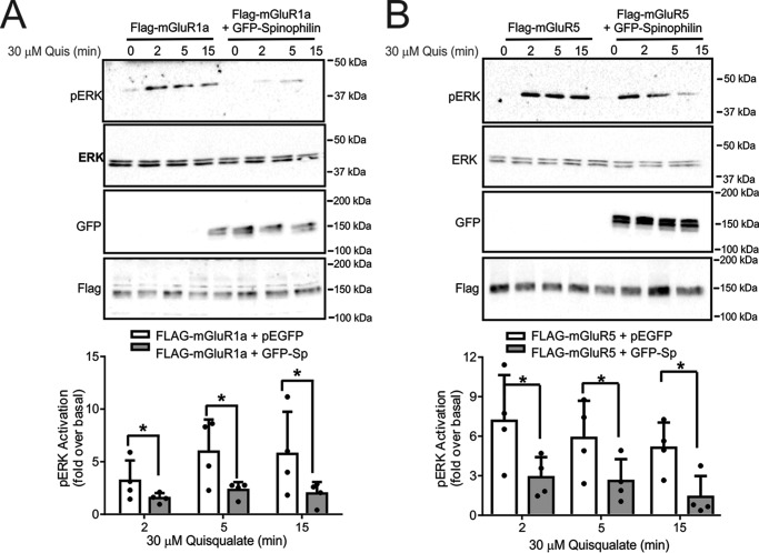
Effect of spinophilin on mGluR1a and mGluR5 ERK1/2 phosphorylation. Shown are representative immunoblots of pERK1/2 activity and total ERK1/2 expression in HEK 293 cells transfected with 2 μg of pcDNA3.1 encoding either FLAG-mGluR1a (A) or FLAG-mGluR5 (B) along with 2 μg of plasmid cDNA encoding either GFP or GFP-spinophilin and treated with 30 μm quisqualate for 0, 2, 5, and 15 min. Also shown are cell lysates (50 μg) for mGluR1a, mGluR5a, and GFP-spinophilin expression. Bar graphs in each panel show the densitometric analysis of ERK1/2 phosphorylation normalized to both basal activity and total ERK1/2 protein expression. Data represent the mean ± S.D. (error bars) of four independent experiments. *, p < 0.05 versus untreated cells lacking GFP-spinophilin.

