
FIGURE 4.
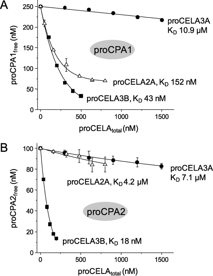
Binding of proCELA2A and proCELA3A to proCPA1 (A) and proCPA2 (B). Equilibrium binding assays using purified proenzymes were carried out as described in the legend to Fig. 3 and under ”Experimental Procedures.“ See text for the error of the fits. For comparison, binding data for wild-type proCELA3B from Fig. 3 are also indicated. Note that KD values in the micromolar range should be considered estimates. The catalytically inactive S217A mutant of proCELA3A was used.