FIGURE 1.

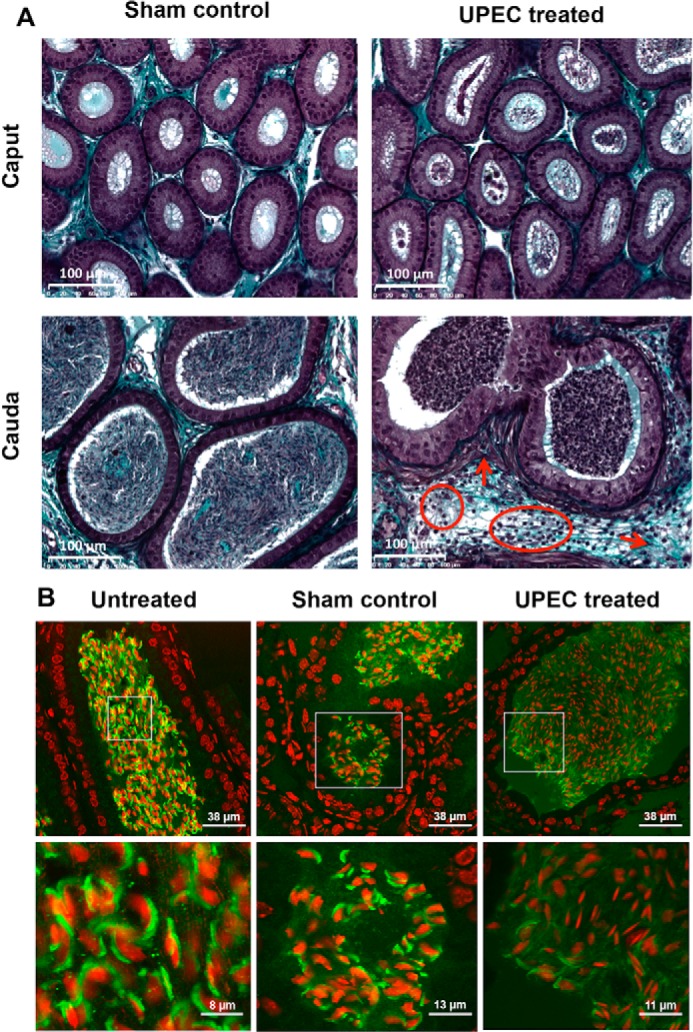
UPEC infection causes pathological alterations in the cauda epididymis and a premature acrosome reaction in epididymal spermatozoa. A, Masson-Goldner-stained sections of cauda epididymal tissues from mice 3 days post-UPEC infection and PBS sham controls were assessed by light microscopy. Arrows indicate fibrotic remodeling (thickening of the smooth muscle cell layer, collagen accumulation). Circles demonstrate interstitial infiltrating leukocytes. B, sperm acrosomes were fluorescently stained with peanut agglutinin-FITC lectin (green), and nuclei were counterstained using TO-PRO-3 (red) in the cauda epididymides from mice 3 days post-UPEC treatment, PBS sham controls, and untreated controls. The insets in the second row were magnified digitally.
