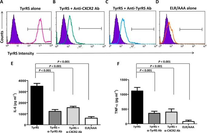
FIGURE 12.
Potential LdTyrRS receptor on mouse macrophages. A–D, FACS analysis to assess the binding of rLdTyrRS to mouse macrophages. rLdTyrRS bound to mouse macrophages was analyzed by indirect staining with the anti-TyrRS antibody. Overlay histograms show binding of rLdTyrRS to the surface of mice macrophages (solid line) with unstained controls (purple solid) as background. Histogram (solid line) shows positive binding of rLdTyrRS to mouse macrophages (A). rLdTyrRS binding to macrophages is reduced when macrophages are preincubated anti-CXCR2 (B) or when rLdTyrRS is neutralized with anti-LdTyrRS antibodies (C). The binding is also significantly reduced with the mutant LdTyrRS (ELR/AAA) protein (D). E and F, cytokine secretion assays for IL-6 (E) and TNF-α (F) using mice macrophages and anti-CXCR2 and anti-LdTyrRS antibodies. The secretion of these proinflammatory cytokines is reduced when macrophages and LdTyrRS protein were preincubated with the anti-CXCR2 (TyrRS + α-CXCR2 Ab) and anti-LdTyrRS (TyrRS + α-TyrRS Ab) antibodies, respectively. The results represent mean ± S.D. with n = 3. Tukey's test was performed, and p values are indicated.

