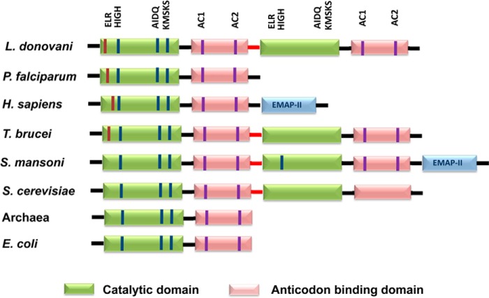
FIGURE 2.
Domain organization of TyrRS from eukaryotes and prokaryotes. The catalytic and anticodon binding domains are indicated. The ELR motif is present at the N terminus and is shown in red. The HIGH and KMSKS active site motifs are common to all class I catalytic domains. The AIDQ motif is characteristic of the ATP binding site in TyrRS. The AC1 motif corresponds to the anticodon-binding domain that interacts with the anticodon stem of tRNATyr. The AC2 motif specifically recognizes the anticodon bases G34 and U/ψ35.

