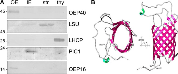
FIGURE 1.
OEP40 forms a β-barrel in the chloroplast OE membrane. A, immunoblot analysis of Ps-OEP40 in chloroplast subfractions. Equal protein amounts (5 μg) of pea chloroplast OE, IE, stroma (str), and thylakoids (thy) were separated by SDS-PAGE and subjected to immunoblot analysis using antibodies directed against recombinant Ps-OEP40. For detection with antisera against marker proteins LSU (str), LHCP (thy), PIC1 (IE), and OEP16.1 (OE), different protein amounts (i.e. 1.0, 0.6, 10.0, and 1.0 μg, respectively) were loaded in each lane. Numbers indicate molecular mass of proteins in kDa. B, three-dimensional structure of a 10-stranded At-OEP40 β-barrel generated by TMBpro. For clarity, the mainly unstructured C terminus starting from His234 (compare supplemental Figs. S1 and S3) is not shown.

