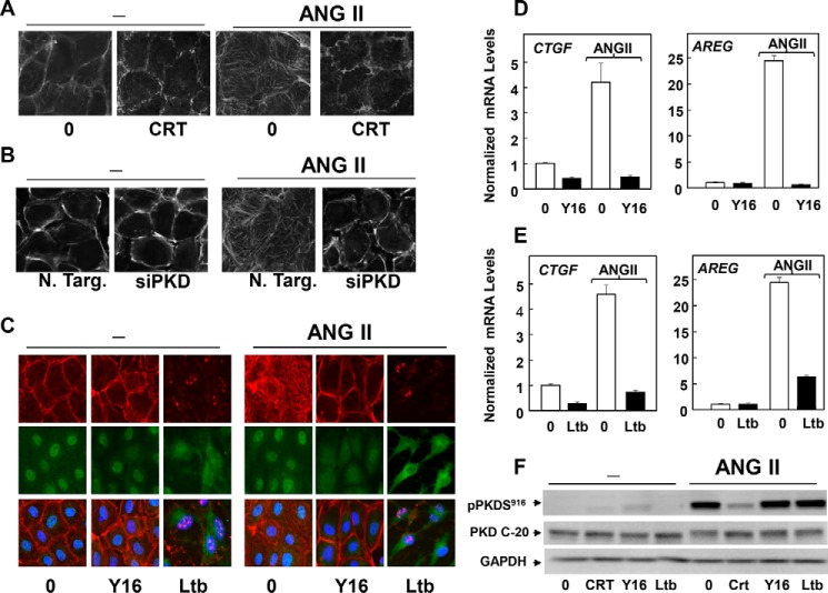
FIGURE 10.
GPCR/PKD mediates YAP re-entry into the nucleus through Rho and actin remodeling. A, confluent and quiescent cultures of IEC-18 cells were incubated in the absence (0) or presence of 2.5 μm CRT0066101 (CRT) for 1 h prior to stimulation without (−) with 10 nm ANG II for 1 h. The cultures were then washed, fixed with 4% paraformaldehyde, and stained with TRITC-conjugated phalloidin. B, cultures of IEC-18 cells were transfected with non-targeting siRNA (N. Targ) or with a mixture of siRNAs targeting PKD1, PKD2, and PKD3 (siPKD) for 4 days. Then the cultures were stimulated with 10 nm ANG II for 1 h, washed, fixed with 4% paraformaldehyde, and stained with TRITC-conjugated phalloidin. C, confluent cultures of IEC-18 cells were incubated in the absence (0) or presence of 20 μm Y16 or 2.5 μm latrunculin B (Ltb) for 1 h prior to stimulation of the cells without (−) or with 10 nm ANG II for 1 h. The cultures were then washed, fixed with 4% paraformaldehyde, and stained with an antibody that detects total YAP, Hoechst 33342, to visualize the cell nuclei and TRITC-conjugated phalloidin. D, confluent cultures of IEC-18 cells were incubated in the absence (0) or presence of 20 μm Y16 for 1 h prior to stimulation of the cells without or with 10 nm ANG II for 1 h, as indicated. RNA was isolated, and the relative levels (n = 3) of Ctgf and Areg mRNA compared with Gapdh mRNA were measured by RT-qPCR. Data are presented as mean ± S.E. Similar results were obtained in three independent experiments. E, confluent cultures of IEC-18 cells were incubated in the absence (0) or presence of 2.5 μm latrunculin B for 1 h prior to stimulation of the cells without or with 10 nm ANG II for 1 h, as indicated. RNA was isolated, and the relative levels (n = 3) of Ctgf and Areg mRNA compared with Gapdh mRNA were measured by RT-qPCR. Data are presented as mean ± S.E. Similar results were obtained in three independent experiments. F, confluent cultures of IEC-18 cells were incubated in the absence (0) or presence of 2.5 μm CRT0066101 (CRT), 20 μm Y16, or 2.5 μm latrunculin B (Ltb) for 1 h prior to stimulation of the cells without (−) or with 10 nm ANG II for 1 h. Cells were then lysed with 2× SDS-PAGE sample buffer and analyzed by immunoblotting antibodies that detect PKD1 phosphorylated at Ser916, total PKD1/2 (PKD C-20), and GAPDH. Similar results were obtained in three independent experiments.

