
FIGURE 2.
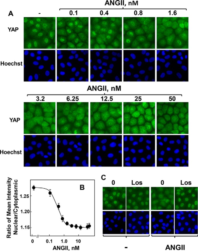
ANG II induces YAP cytoplasmic localization in a dose-dependent manner via the AT1 receptor in IEC-18 cells. A, confluent cultures of IEC-18 cells were stimulated with ANG II at the indicated concentrations for 30 min. The cultures were then washed, fixed with 4% paraformaldehyde, and stained with an antibody that detects total YAP and with Hoechst 33342 to visualize the cell nuclei. B, quantification of nuclear/cytoplasmic ratio of YAP immunofluorescence shown in A was determined with the CellProfiler software as described under “Experimental Procedures.” The plot shown are the mean ratios ± S.E. n = 7 fields (∼1250 cells were analyzed for each concentration of ANG II). Similar results were obtained in a separate experiment. C, confluent cultures of IEC-18 cells were incubated without (0) or with the selective ANG II type 1 (AT1) receptor antagonist losartan (Los) at 10 μm for 30 min prior to stimulation with 10 nm ANG II for 30 min. The cultures were then washed, fixed with 4% paraformaldehyde, and stained with an antibody that detects total YAP and with Hoechst 33342 to visualize the cell nuclei. Similar results were obtained in three independent experiments.