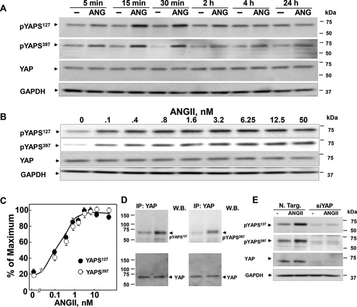
FIGURE 3.
ANG II induces rapid increase in the phosphorylation of YAP at Ser127 and Ser397 in IEC-18 cells. A and B, confluent cultures of IEC-18 cells were incubated without (−) or with ANG II (ANG) for the indicated times (A) or concentrations (B). Cultures were then lysed with 2× SDS-PAGE sample buffer and analyzed by immunoblotting with antibodies that detect YAP phosphorylated at Ser127 and Ser397. Immunoblotting for total YAP and GADPH was also included. C, quantification of total YAP phosphorylated at Ser127 and Ser397 was performed using MultiGauge version 3.0. The results represent the mean ± S.E., n = 3, and are expressed as percentage of the maximal level of YAP phosphorylated at Ser127 and Ser397. Similar results were obtained in three independent experiments. D, confluent cultures of IEC-18 cells were stimulated with 10 nm ANG II for 30 min. Then the cells were lysed, and YAP immunoprecipitates (IP: YAP) were analyzed by Western blotting (W.B.) with YAP Ser127 and Ser397. E, cultures of IEC-18 cells were transfected with non-targeting siRNA (N. Targ) or with siRNAs targeting YAP for 4 days. The cultures were then stimulated with 10 nm ANG II for 30 min. Cells were lysed with 2× SDS-PAGE sample buffer and analyzed by immunoblotting antibodies that detect YAP phosphorylated at Ser127 and Ser397, total YAP, and GAPDH. Similar results were obtained in four independent experiments.

