FIGURE 5.
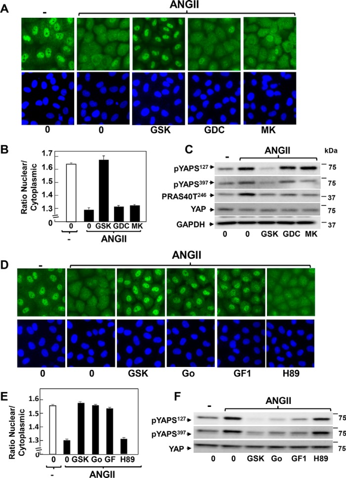
ANG II induces cytoplasmic localization and phosphorylation of YAP through an Akt-independent but PKC-dependent pathway in IEC-18 cells. A, confluent cultures of IEC-18 cells were incubated in the absence (0) or presence of 2.5 μm GSK690693 (GSK), 2.5 μm GDC-0068 (GDC), or 5 μm MK2206 (MK) for 1 h prior to stimulation of the cells without (−) or with 10 nm ANG II for 30 min. The cultures were then washed, fixed with 4% paraformaldehyde, and stained with an antibody that detects total YAP and with Hoechst 33342 to visualize the cell nuclei. B, quantification of the nuclear/cytoplasmic ratio of YAP immunofluorescence shown in A was determined with the CellProfiler software. The bars shown are the mean nuclear/cytoplasmic ratio ± S.E. n = 6 fields (∼1,200 cells were analyzed for each condition). Similar results were obtained in three independent experiments. C, confluent cultures of IEC-18 cells were incubated in the absence (0) or presence of 2.5 μm GSK690693 (GSK), 2.5 μm GDC-0068 (GDC), or 5 μm MK2206 (MK) for 1 h prior to stimulation of the cells without (−) or with 10 nm ANG II for 30 min. Cells were then lysed with 2× SDS-PAGE sample buffer and analyzed by immunoblotting with antibodies that detect YAP phosphorylated at either Ser127 or Ser397, PRAS40 phosphorylated at Thr246, and total YAP and GADPH. Similar results were obtained in three independent experiments. D, confluent cultures of IEC-18 cells were incubated in the absence (0) or presence of 2.5 μm GSK690693 (GSK), 5 μm Gö6983 (Go), 5 μm GF1, or 10 μm H89 for 1 h prior to stimulation of the cells without (−) or with 10 nm ANG II for 30 min. The cultures were then washed, fixed with 4% paraformaldehyde, and stained with an antibody that detects total YAP and with Hoechst 33342 to visualize the cell nuclei. E, quantification of the nuclear/cytoplasmic ratio of YAP immunofluorescence shown in D was determined with the CellProfiler software. The bars shown are the mean nuclear/cytoplasmic ratios ± S.E. n = 6 fields (∼1,100 cells were analyzed for each condition). Similar results were obtained in four independent experiments. F, confluent cultures of IEC-18 cells were incubated in the absence (0) or presence of 2.5 μm GSK690693 (GSK), 5 μm Gö6983 (Go), 5 μm GF1, or 10 μm H89 for 1 h prior to stimulation of the cells without (−) or with 10 nm ANG II for 30 min. Cells were then lysed with 2× SDS-PAGE sample buffer and analyzed by immunoblotting antibodies that detect YAP phosphorylated at Ser127 and Ser397 and total YAP. Similar results were obtained in three independent experiments.

