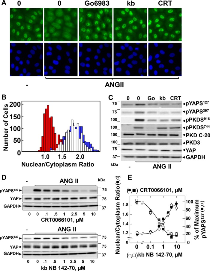
FIGURE 6.
PKD family inhibitors prevent transient YAP translocation and phosphorylation in response to ANG II in IEC-18 cells. A, confluent cultures of IEC-18 cells were incubated in the absence (0) or presence of 5 μm Gö6983, 3.5 μm kb NB 142-70 (kb), or 2.5 μm CRT0066101 (CRT) for 1 h prior to stimulation of the cells without (−) or with 10 nm ANG II for 30 min. The cultures were then washed, fixed with 4% paraformaldehyde, and stained with an antibody that detects total YAP and with Hoechst 33342 to visualize the cell nuclei. B, quantification of the nuclear/cytoplasmic ratio of YAP immunofluorescence was determined using CellProfiler. The bars shown represent control (open bars), ANG II (red bars), ANG II + CRT (blue bars) and are the nuclear/cytoplasmic ratios from ∼1,000 cells from one experiment. Similar results were obtained in six independent experiments. C, confluent cultures of IEC-18 cells were incubated in the absence (0) or presence of 5 μm Gö6983 (Go), 3.5 μm kb NB 142-70 (kb), or 2.5 μm CRT0066101 (CRT) for 1 h prior to stimulation of the cells without (−) or with 10 nm ANG II for 30 min. Cells were then lysed with 2× SDS-PAGE sample buffer and analyzed by immunoblotting antibodies that detect YAP phosphorylated at Ser127 and Ser397, PKD1 phosphorylated at Ser916, PKDs phosphorylated at Ser744, and total PKD1/2 (PKD C-20), PKD3, YAP, and GAPDH. Similar results were obtained in six independent experiments. D and E, confluent cultures of IEC-18 cells were incubated in the absence (0) or presence of increasing concentrations of either CRT0066101 (closed squares, in E) or kb NB 142-70 (open squares in E) for 1 h prior to stimulation of the cells without (−) or with 10 nm ANG II for 30 min. Cells were then lysed with 2× SDS-PAGE sample buffer and analyzed by immunoblotting antibodies that detect YAP phosphorylated at Ser127, total YAP, and GAPDH. Quantification YAP Ser127 phosphorylation (shown in E) was performed using MultiGauge version 3.0. The results represent the mean ± S.E., n = 3, and are expressed as percentage of the maximum level of YAP Ser127. E, confluent cultures of IEC-18 cells incubated in the absence (gray circle) or presence of increasing concentrations of CRT0066101 (closed circles) or kb NB 142-70 (open circles) for 1 h as indicated prior to stimulation of the cells without or with 10 nm ANG II for 30 min. The cultures were then washed, fixed with 4% paraformaldehyde, and stained with an antibody that detects total YAP and with Hoechst 33342 to visualize the cell nuclei and analyzed with the CellProfiler software. The results shown are the nuclear/cytoplasmic ratio of YAP immunofluorescence ± S.E.; ∼700 cells were analyzed at each concentration of CRT0066101 (closed circles) or kb NB 142-70 (open circles). Similar results were obtained in two independent experiments.

