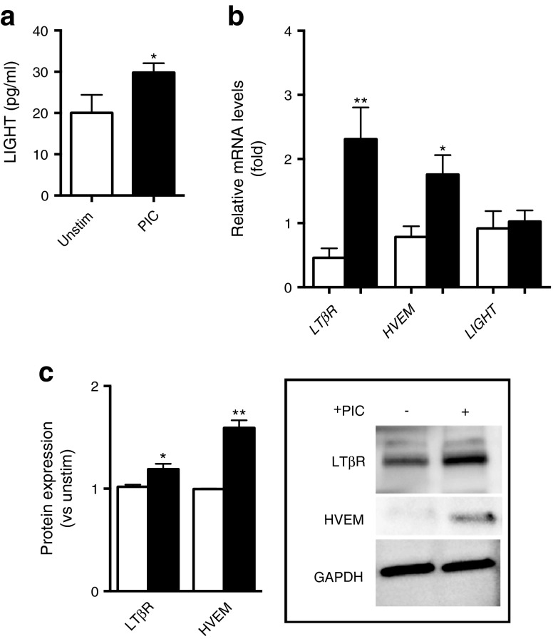
Fig. 3.
Proinflammatory stimuli increase the expression of LIGHT in human islets. Islets from independent preparations were stimulated for 24 h (a, b) or 48 h (c) with (black bars) or without (white bars) a PIC (IL-1β [1 ng/ml], IFN-γ [50 ng/ml], and TNF [10 ng/ml]) before the levels of LIGHT (ng/ml) were determined by ELISA in cell supernatant fractions (a). (b) Expression of the LIGHT receptors (LTβR and HVEM) and LIGHT mRNA levels as assessed by quantitative PCR in relation to the control gene β actin. (c) Protein levels of the LIGHT receptors in relation to the protein expression levels of GAPDH as assessed by western blotting. Data are presented as mean ± SEM (n = 3–6). *p < 0.05 and **p < 0.01 vs unstimulated cells using a Mann–Whitney U test. Unstim, unstimulated

