FIGURE 5.
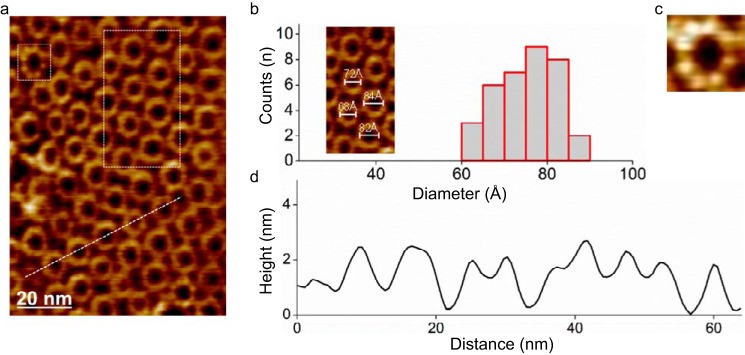
Visualization of pores of WT FraC with AFM. a, two-dimensional packing of ring-shaped oligomers of WT FraC on supported lipid bilayers composed of the lipid mixture SM/DOPC (1:1). b, diameter distribution analysis (peak-to-peak distances of the protein protrusion in the height profile). The average diameter of the particles was 75 ± 6 Å (mean ± S.D. from the Gaussian distributions). Inset, detail of the particles inside the white dashed rectangle in a. c, magnification (13-nm frame size) of a single FraC oligomer in a (white dashed square). d, cross-section profile (left to right) of FraC oligomers shown in a (white dashed line). The molecules are packed with a center-to-center distance of ∼112 Å.

