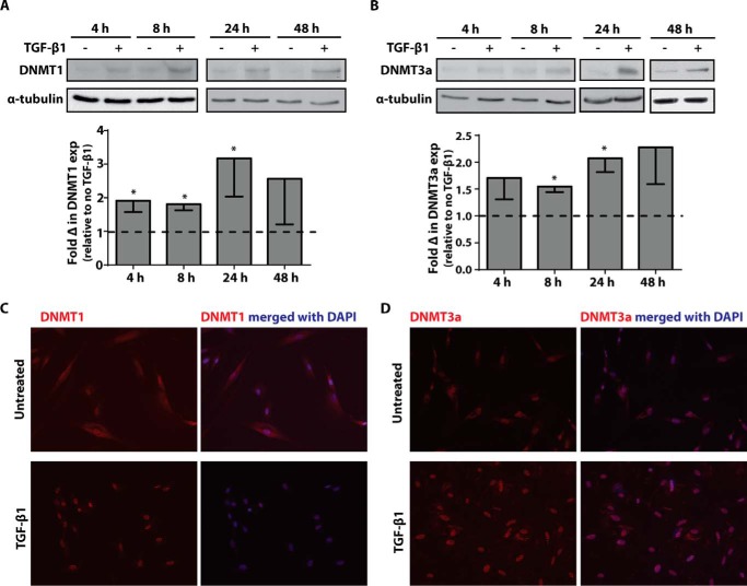
FIGURE 1.
Treatment with TGF-β1 increased DNMT1 and DNMT3a protein expression. A and B, CCL210 cells were treated with 2 ng/ml of TGF-β1 for 4, 8, 24, and 48 h, and DNMT1 (A) and DNMT3a (B) protein expression was assayed by immunoblot. A representative immunoblot is shown with the mean fold change in expression by densitometry, relative to no-TGF-β1 control, shown below (n = 4). The data are shown as means ± S.E. * p < 0.05 relative to no-TGF-β1 treatment. C and D, cells were treated with or without TGF-β1 (2 ng/ml) for 8 h and immunostained for DNMT1 (C) and DNMT3a (D) with a TRITC-conjugated secondary antibody. Nuclei were counterstained with DAPI.

