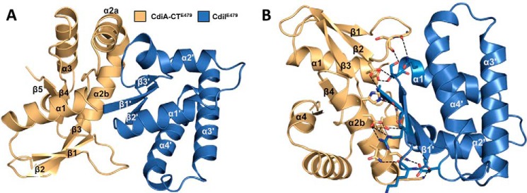
FIGURE 1.
Structure of the CdiA-CT/CdiIE479 toxin/immunity protein complex. A, CdiA-CT/CdiIE479 complex is shown as a schematic with secondary structure elements of CdiI indicated with prime symbols. B, CdiA-CT/CdiIE479 complex formation is mediated by electrostatic interactions. Interacting residues are in stick representation with nitrogen and oxygen atoms colored in blue and red (respectively), and bonds are shown as black dashed lines. The view is rotated 180° about the x axis relative to A.

