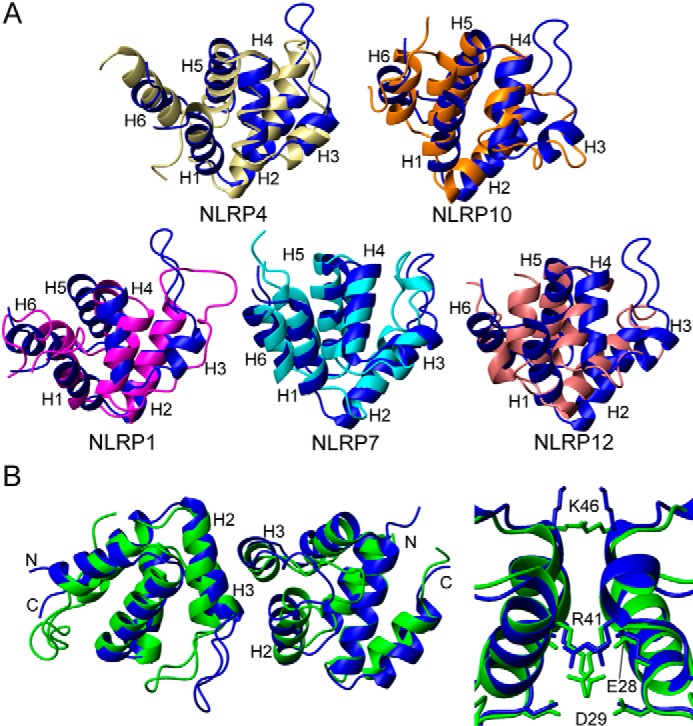
FIGURE 7.

Comparison of NLRP3 PYD with other pyrin domains. A, superposition of the known three-dimensional solution structures of NLRP pyrin domains to NLRP3 PYD represented as ribbon diagrams (PDB codes: NLRP3, 2NAQ; NLRP1, 1PN5; NLRP4, 4EWI; NLRP7, 2KM6; NLRP10, 2DO9; and NLRP12, 2l6A). Blue (NLRP3), magenta (NLRP1), ivory (NLRP4), cyan (NLRP7), orange (NLRP10), and pink (NLRP12) are shown. Helices are numbered. Superimposed residues belong to helices 1, 2, and 4–6. Helix 3 (absent in NLRP1, NLRP10, and NLRP12) and the loop connecting helices 2 and 3 were excluded because of significant conformational variability. The r.m.s.d. values between NLRP3 and the other PYDs are as follows: NLRP4, 3.9 Å; NLRP10, 3.4 Å; NLRP1, 4.9 Å; NLRP7, 5.2 Å; and NLRP12, 5.9 Å. B, superposition of the crystallographic dimeric structure of NLRP3 PYD (green, PDB code, 3QF2; Ref. 36) onto the monomeric NMR structure (blue). The image at the right side shows a detailed view of the dimeric interface, including side chain orientation. The image was displayed with MOLMOL (65).
