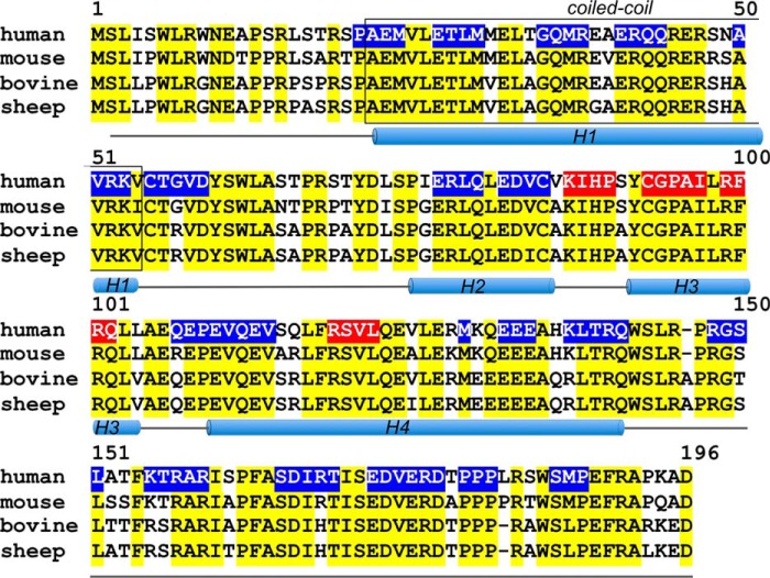
FIGURE 3.
The primary structure of a human RD3 aligned with the mouse, bovine, and sheep protein sequences. Positions of the identical residues are marked in yellow, the H1–H4 regions are predicted α-helices, the boxed region is a predicted coiled-coil (after Molday et al. (8)), and the line marks regions lacking a defined secondary structure. The regions where scrambling of the peptide sequences reduced RetGC1 inhibition by RD3 by ≤50% (see Fig. 4) are highlighted in blue; those where the mutation reduced the extent of inhibition by ≥80% are highlighted in red.

