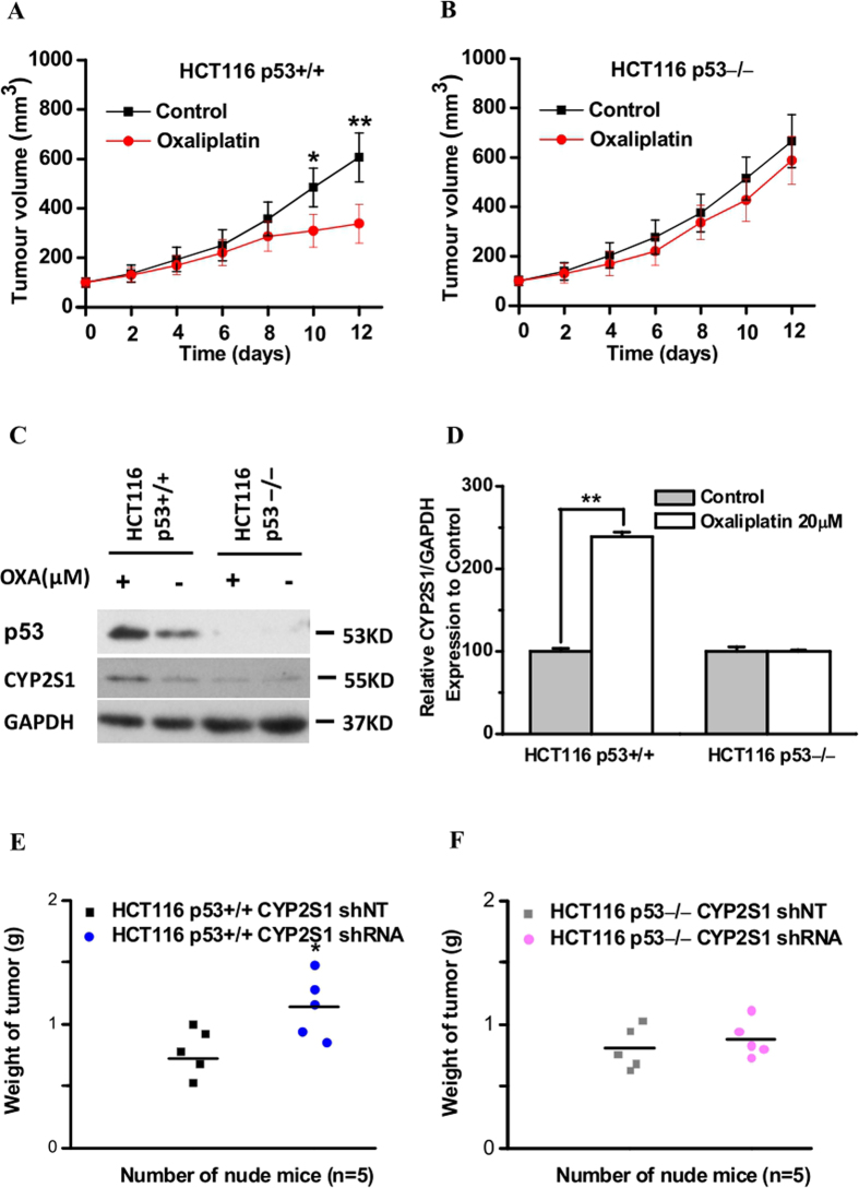
Figure 5. Upregulation of CYP2S1 protein expression increases oxaliplatin sensitivity in a p53-dependent manner in vivo.
Identical (2 × 10⁶) amounts of each cells were injected subcutaneously into the flanks of nude mice. When tumors reached approximately 100 mm3, the mice received either PBS or oxaliplatin (10 mg/kg) intraperitoneally (day 0). A second dose of either PBS or oxaliplatin was administered three days later. Tumor growth was analyzed by caliper measurements every 2 days. (A,B) Comparison of tumor volume between each group (means ± SD, n = 5). Oxaliplatin treatment markedly reduced tumor volume in p53+/+HCT116 tumor xenografts. *p < 0.05, **p < 0.01. (C) Tumors were harvested on day 12. Tumors from each group were analyzed for the presence of CYP2S1 and p53 by western blotting. GAPDH was used as an internal control. (D) Total proteins were extracted and the protein levels of the CYP2S1 were analyzed in Western blot analysis. Results are of the average from three experiments. Data are represented as mean ± s.d., ** p < 0.01. (E,F) Weights of xenograft tumours derived from p53+/+ or p53−/−HCT116 cells expressing CYP2S1 knockdown or control (shRNA or shNT, respectively) after oxaliplatin treatment. n = 5 mice per group.

