
Figure 4. A schematic explaining the image formation of a charged region on the screen.
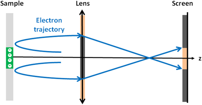
The electron beam is deviated by a negatively charged region on the surface of the sample. In a direct image, the region appears as a large dark spot on the screen. In the presence of a converging lens, the electron beam becomes narrower on the screen and the negatively charge region appears as a small bright spot. Adapted from ref. 37.