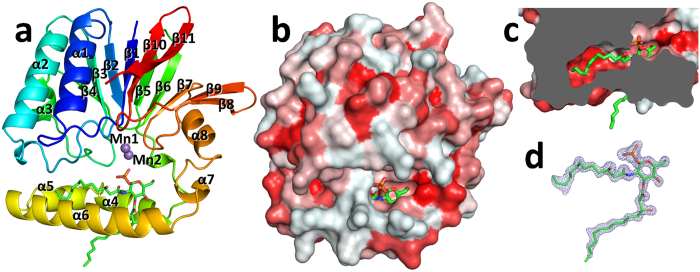
Figure 2. Structure of PaLpxH.
(a) Ribbon diagram of the Pseudomonas aeruginosa LpxH (PaLpxH) shown in rainbow color (P21 crystal with Mn2+). Lipid X is depicted by green sticks and the Mn2+ ions are violet spheres. PaLpxH consists of a catalytic domain of approximately 180 residues (Met1–Leu118 and Val174–Leu240) and a helical insertion domain (HI domain) (α4–α7). (b) Surface representation of the PaLpxH. The structure is colored in red with different density based on the hydrophobicity scale30. (c) Cross-sectional view of the PaLpxH focusing to hydrophobic cavity between the catalytic and HI domains. The 2-acyl chain of the lipid X is deeply buried in the cavity. d. Fo–Fc omit maps superposed with bound lipid X (cut off 3.5 σ).

