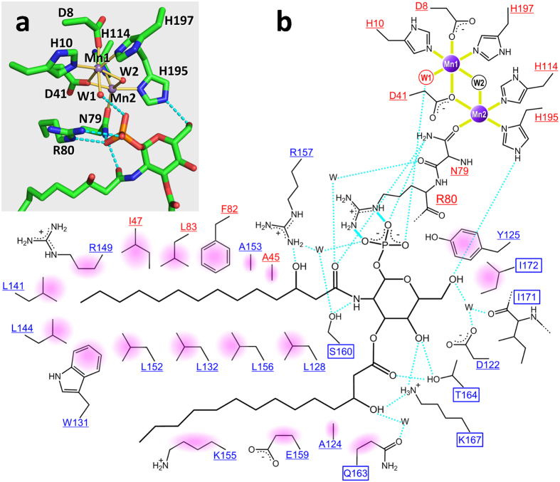
Figure 3. Detailed representation of Mn2+ and lipid X recognition by PaLpxH.
(a) Residues involved in Mn2+ coordination and the binding of the glucosamine-1-phosphate moiety of lipid X are shown. Mn2+ coordination is depicted with yellow bonds and polar interactions are depicted with light blue dotted lines. Mn1 is in octahedral coordination with six ligands, whereas Mn2 has five ligands with one open site facing the phosphate group of lipid X. Water molecules (W1 and W2) are shown as red spheres. (b) Schematic overview of Mn2+ and lipid X binding. Residues in the catalytic domain are shown in red and those in the HI domain are shown in blue. Residues whose structures change upon lipid X binding are highlighted in squares, whereas residues whose structures are unchanged are underlined. Coordination bonds are indicated with solid yellow lines, bidentate salt bridges are indicated with light blue solid lines, and hydrogen bonds are indicated with light blue dotted lines. Apolar interactions are shown with pink shading.

