Fig. 6.

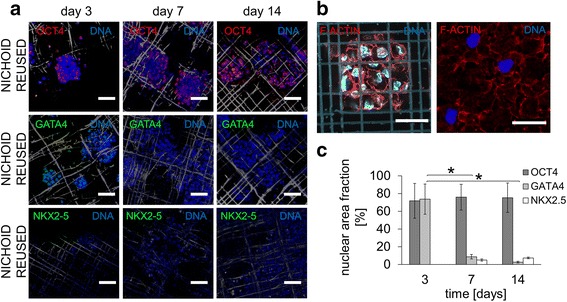
Spontaneous differentiation of mES cells cultured on reused nichoid substrates. Cells were cultured in the absence of a feeder layer and with LIF up to day 3, then without either a feeder layer or LIF from days 4 to 14. a Immunofluorescence for OCT4 (red), GATA4 (green), NKX2.5 (green) and DAPI (blue) at days 3, 7, and 14. The scale bar is 50 μm. b Immunofluorescence for F-actin (red) and DAPI (blue) showing a few residual embryoid bodies anchored both to the nichoid (left) and the flat glass (right) after trypsinization. The scale bar is 30 μm. c Quantification of OCT4, GATA4 and NKX2.5 expression by image processing; n = 15, * p < 0.01
