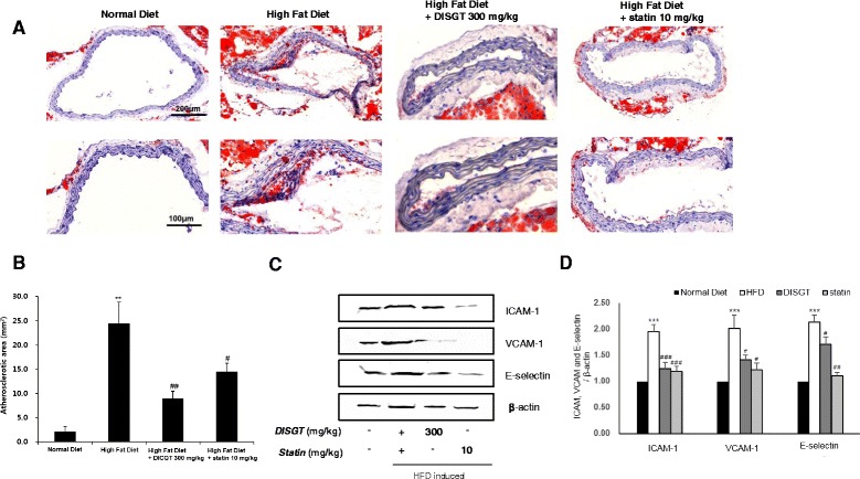
Fig. 2.

Effect of DISGT on reducing aortic lesions and expression of adhesion molecules in the aorta. a The aorta was collected from HFD-fed mice that were treated with DISGT or statin orally for 16 weeks. Representative photomicrographs of Oil Red O staining are shown. (Cross sections; ×100, above; ×200, under, magnification). b Quantification of Oil Red O-stained atherosclerotic lesion areas from each group. c Western blots of ICAM-1, VCAM-1, and E-selectin. d Densitometric analysis showing the relative protein expression levels of ICAM-1, VCAM-1, and E-selectin that were normalized to β-actin. Values are mean ± SEM. **p < 0.01, ***p < 0.001 vs. ND; # p < 0.05, ## p < 0.01, ### p < 0.001vs. HFD
