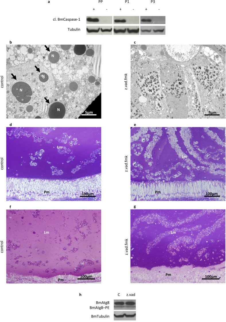
Figure 8. Caspase inhibition delays the degeneration of the midgut epithelium.
(a) Western blot analysis of cleaved BmCaspase-1 in midgut of larvae treated with z.vad.fmk. The absence of activated caspase expression can be appreciated at PP, P1, and P3 stages following the administration of the inhibitor at SD2 stage; (b,c) TEM analysis of midgut in control (b) and treated (c) larvae at PP stage; (d,e) morphology of larval and pupal epithelium in control (d) and treated (e) P3 pupae; (f,g) morphology of larval and pupal epithelium in control (f) and treated (g) P9 pupae; (h) Western blot analysis of BmAtg8–PE in the midgut of z.vad.fmk-treated larvae. Lm: larval midgut epithelium; N: nucleus; Pm: pupal midgut epithelium; arrows: apoptotic nuclei.

