
Fig. 1.
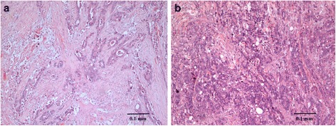
a Cancerous tissue section of patients Dukes' B well-differentiated adenocarcinoma. Hematoxylin (purple) stains chromatin in the nucleus and eosin (pink orangish) gives color to the protein that resides in the cytoplasm of muscle cells. Tumor cells appear to thicken and be seen spreading muscular propia but did not penetrate serous layer. b. Well differentiated adenocarcinoma Dukes’ C tissue section invaded into muscular propia and involved lymph nodes