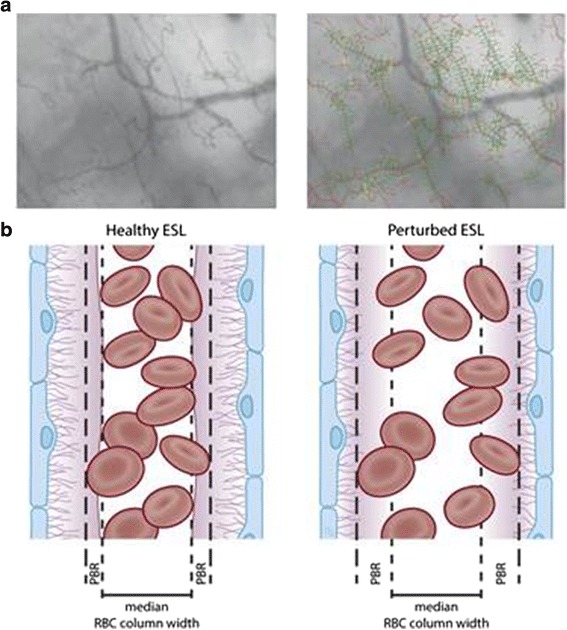
Fig. 5.

Sidestream dark field (SDF) imaging for measuring the perfused boundary region (PBR) in the sublingual capillary bed. a Recording of the sublingual capillary bed captured using an SDF camera (left). The capillaries are automatically recognized and analyzed after various quality checks (right). Based on the shift in the red blood cell (RBC) column width over time, the PBR can be calculated. b Model of a blood vessel showing the PBR under healthy conditions (left). The EG prevents the RBC from approaching the endothelial cell; thus, the PBR is relatively small. Under disease conditions (right) or after enzymatic breakdown of the EG in an animal model, the damaged EG allows the RBCs to approach the endothelium more often. This results in a higher variation in RBC column width, which is reflected as a high PBR. ESL, endothelial surface layer (cited from Dane MJ, van den Berg BM, et al. Am J Physiol Renal Physiol. 2015,308(9):F956–F966)
