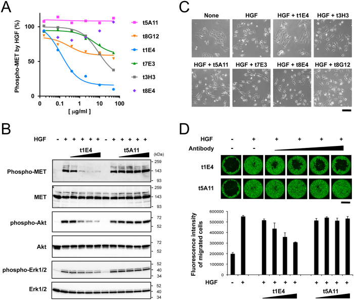
Figure 2. Effect of anti-HGF antibodies on the HGF-induced cellular responses.
(A) Effect of anti-HGF antibodies on the HGF-induced cellular Met activation. The EHMES-1 cells were stimulated with 20 ng/ml HGF in the presence of vairious antibodies at indicated concentrations for 10 min. The phospho-Met level was determined as in Fig. 1 and expressed as the relative value obtained in the absence of antibody. Data are from a representative experiment in which triplicate determinations were made. (B) Inhibition of HGF-induced phosphorylation of Met, Akt, and ERK by t1E4. The EHMES-1 cells were stimulated for 10 min with (+) or without (−) 20 ng/ml HGF, together with the increasing concentrations (0.08, 0.4, 2, and 10 μg/ml) of t1E4 or t5A11 IgGs. Cell lysates were analyzed by SDS-PAGE and Western blotting. (C) Effect of anti-HGF antibodies on the ability of HGF to induce MDCK cell scattering. MDCK cells were left untreated (None) or stimulated with 2 ng/ml HGF for 16 h, in the presence of indicated antibodies at 10 μg/ml. Scale bar, 100 μm. (D) Inhibition of the HGF-stimulated migration of HuCCT1 human liver bile duct carcinoma cells by t1E4. Images of migrated HuCCT1 cells stimulated with (+) or without (−) 20 ng/ml HGF in the presence of increasing concentrations (0.08, 0.4, 2, and 10 μg/ml) of t1E4 or t5A11 for 24 hrs (upper panels). Cells were stained by calcein-AM. Scale bar, 1 mm. Migrated cells stained with calcein-AM were quantified by fluorescence intensity (lower graph). Data are from a representative experiment in which triplicate determinations were made.

