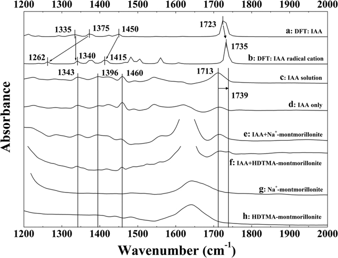
Figure 5.

Comparison of calculated and experimental IR spectra: calculated IR spectra of (a) 3-indole-acetic-acid and (b) 3-indole-acetic-acid radical cation; observed IR spectra of (c) 3-indole-acetic-acid in solution, (d) 3-indole-acetic-acid solution irradiated by mercury lamp, and 3-indole-acetic-acid solution irradiated by mercury lamp in the presence of (e) Na+-montmorillonite, (f) HDTMA-montmorillonite; observed IR spectra of (g) Na+-montmorillonite, (h) HDTMA-montmorillonite irradiated by mercury lamp. Experimental conditions: the initial concentrations of 3-indole-acetic-acid, and clay mineral were 10 mM and 5 g L−1, respectively; pH was adjusted to 4.0 by adding NaOH and HCl; an arc light source equipped with a 350 W mercury lamp was used as the light source.
