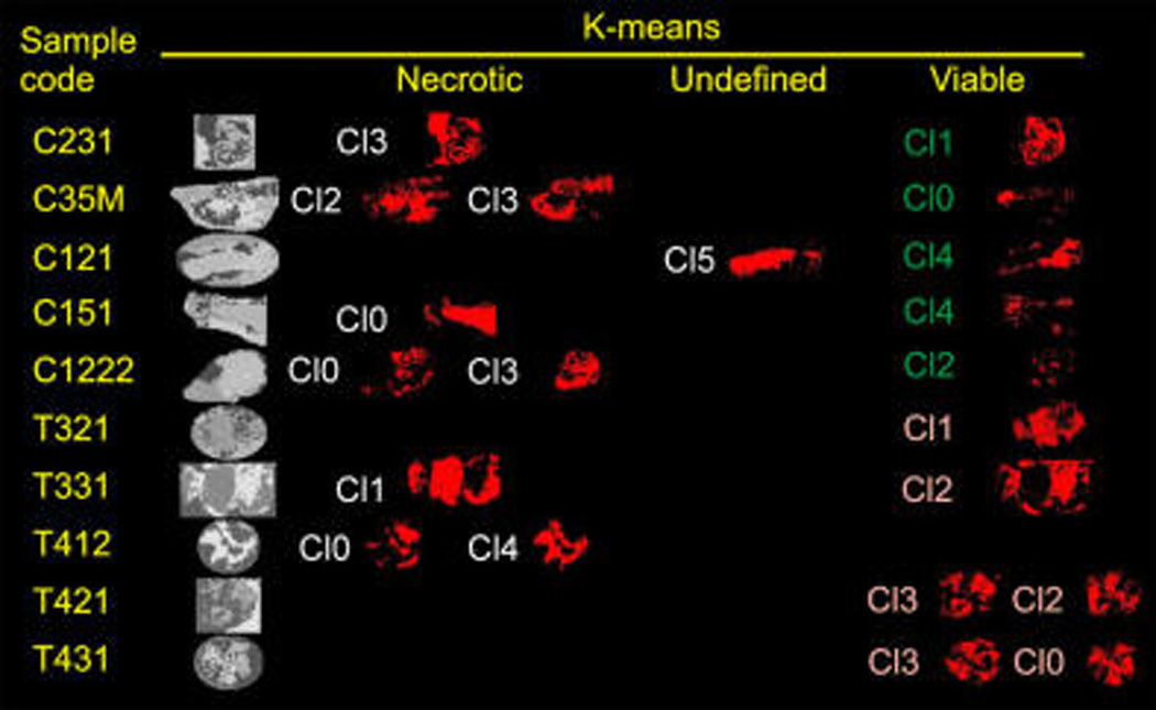
Figure 1.

k-means analysis of eight sections of control (C) and treated (T) xenografts: gray scale images, ROIs identified by the algorithm in each section; necrotic, regions identified as containing mainly necrotic tissue; viable, regions identified as containing mainly viable tissue; undefined, sections in which necrotic or viable tissue was not clearly grouped.
