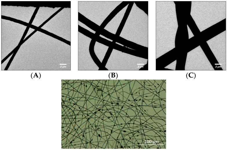
Figure 1.
(Top) TEM image of (A) 12.5% w/v (B) 15% w/v and (C) 17.5% w/v PS microfibres. Fibres were spun onto carbon-coated grids for 15 s. Micrographs taken using a type CM 12 Philips TEM at 120 kV; (Bottom) Morphology of 12.5% w/v PS microfibres. Fibres were spun onto a metal collector plate and transferred to a UVO-treated piece of PMMA. Image taken with a Nikon Digital Eclipse C1 confocal microscope in brightfield setting.

