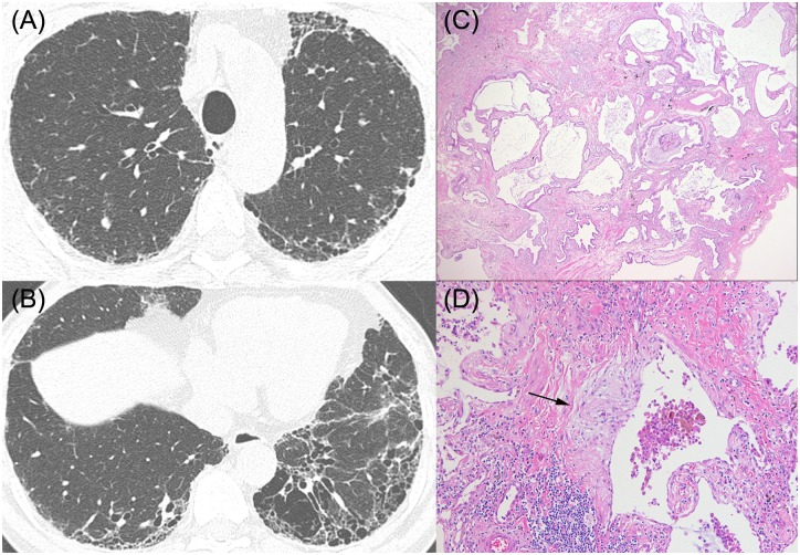
Fig 5. HRCT images and pathologic features of a 66-year-old man who was categorized as UIP without emphysema.
(A), (B) HRCT shows subpleural honeycombing predominantly in the lower lobes without emphysema (C), (D) Pathologic features show dense fibrosis with architectural distortion, microscopic honeycomb change (C, x20), and often fibroblastic foci (arrow), (D, x200). He was considered to have idiopathic pulmonary fibrosis, and pSRIF score was -2.

