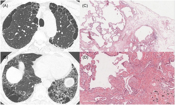
Fig 7. HRCT images and pathologic features of a 67-year-old man who was diagnosed with SRIF in pathology.
(A), (B) HRCT shows upper lobe-predominant paraseptal emphysema, and asymmetric, inhomogeneous honeycombing with bullae in the lower lobes. (C, D) Pathologic features show emphysematous change, interstitial fibrosis and dense fibrosis with architectural distortion and fibroblastic focus (x20, x40). He was diagnosed with SRIF by multidisciplinary discussion, because also he was not suspicious to have IPF in clinical information. The pSRIF score was 2.

