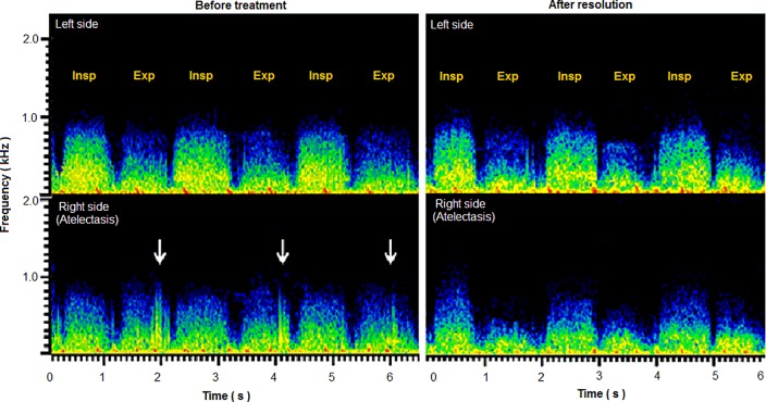
Fig 2. Representative sound spectrograms in a child with right middle lobe atelectasis before and after treatment.
The sound spectrograms for recordings over bilateral middle lung fields are shown. Before treatment, the inspiratory sound intensity was lower on the right than on the left. Furthermore, on the right (affected) side, the expiratory sound intensity was similar to the inspiratory sound intensity. In addition, coarse crackles (white arrows) were identified. After resolution, the right-to-left difference in inspiratory sound intensity persisted. On the other hand, the recovery of a normal pattern of inspiratory breath sound dominance over expiratory breath sound was observed and the adventitious sounds disappeared.

