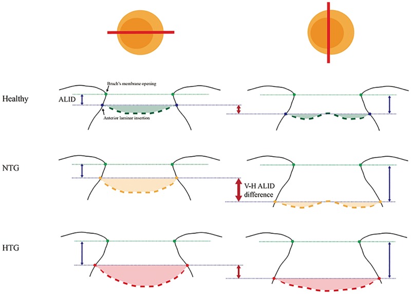
Fig 4. Schematic diagram showing LC-structural differences among HTG, NTG and healthy eyes.
In healthy eyes, the anterior laminar insertion locates more posteriorly in superior and inferior axis compared to nasal and temporal axis, which results in vertical-horizontal ALID difference (small red arrow in the first line). In NTG eye, the vertical (superior-inferior) LC insertion locates much deeper than horizontal (nasal-temporal) LC insertion, which lead to increased vertical-horizontal ALID difference (large, thicker red arrow in the second line). The laminar curvature is increased in both meridians (yellow shaded area). In HTG eye, the horizontal anterior laminar insertion further locates posteriorly, which results in decreased vertical-horizontal ALID difference. The laminar curvature is much increased, so that the ‘w-shape’ contour changes to ‘u-shape’ contour in vertical meridian. The green points indicate Bruch’s membrane opening, and the thin green dotted-line corresponds to the reference plane connecting the BMO. The thicker green (healthy), yellow (NTG), and orange (HTG) dotted-lines indicate the anterior LC.

