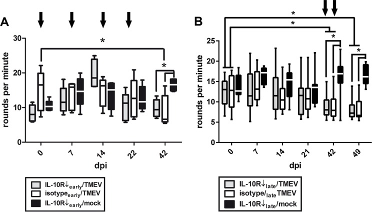
Fig 2. Motor coordination in Theiler’s murine encephalomyelitis virus (TMEV)-infected SJL mice.
(A) Experiment I: RotaRod® performance test after early application of the IL-10 receptor blocking antibody (IL-10R Ab) at 0, 7, 14 and 21 dpi (arrows) revealed motor coordination deficits in TMEV-infected animals compared to mock-infected mice at 42 dpi. However, no differences were determined between IL-10R Ab- and isotype-treated animals. white box = TMEV-infected mice without IL-10R Ab treatment (group “isotypeearly/TMEV”), grey box = TMEV-infected mice with IL-10R treatment (group “IL10R↓early/TMEV”), black box = mock-infected mice with IL-10R treatment (group “IL10R↓early/mock”). (B) Experiment II: application of IL-10R Ab at 35 and 42 dpi (arrows) caused also deterioration of motor coordination starting at 42 dpi. However, no differences were observed between IL-10R Ab- and isotype-treated animals (groups “IL-10R↓late/TMEV” and “isotypelate/TMEV”). white box = TMEV-infected mice without IL-10R Ab treatment (group “isotypelate/TMEV”), grey box = TMEV-infected mice with IL-10R treatment (group “IL10R↓late/TMEV”), black box = mock-infected mice with IL-10R treatment (group “IL10R↓late/mock”). Box and whisker plots display median, minimum and maximum values as well as upper and lower quartiles, 5 animals used at all investigated time points, Wilcoxon rank-sum tests, * = p < 0.05.

