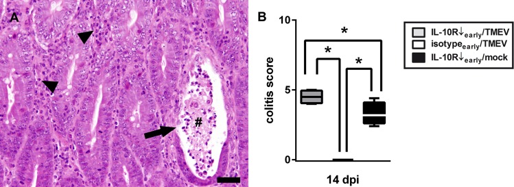
Fig 7. Theiler’s murine encephalomyelitis virus (TMEV)-infection exacerbates enteric disease following interleukin-10 receptor (IL-10R) blockade.
(A) Note changes of colonic mucosa with severe lymphohistiocytic to neutrophilic inflammation (arrow heads), crypt abscess formation (#), and loss of crypt epithelium (arrow). H&E staining, bar = 60 μm (B) Significantly increased severity of colitis at 14 days post infection (dpi) in TMEV-infected mice with IL10R antibody (Ab) treatment (group “IL10R↓early/TMEV”) compared to Ab treated animals without infection (group “IL10R↓early/mock”). Animals receiving isotype control instead of IL-10R Ab did not show any intestinal inflammation. IL-10R Ab treated SJL mice with TMEV-infection (group “IL10R↓early/TMEV”), TMEV-infected mice without IL-10R Ab treatment (group “isotypeearly/TMEV”), IL-10R Ab treated SJL mice without TMEV-infection (group “IL10R↓early/mock”). Box and whisker plots display median, minimum and maximum values as well as upper and lower quartiles, 5 animals used in all groups, Wilcoxon rank-sum tests, * = p < 0.05.

