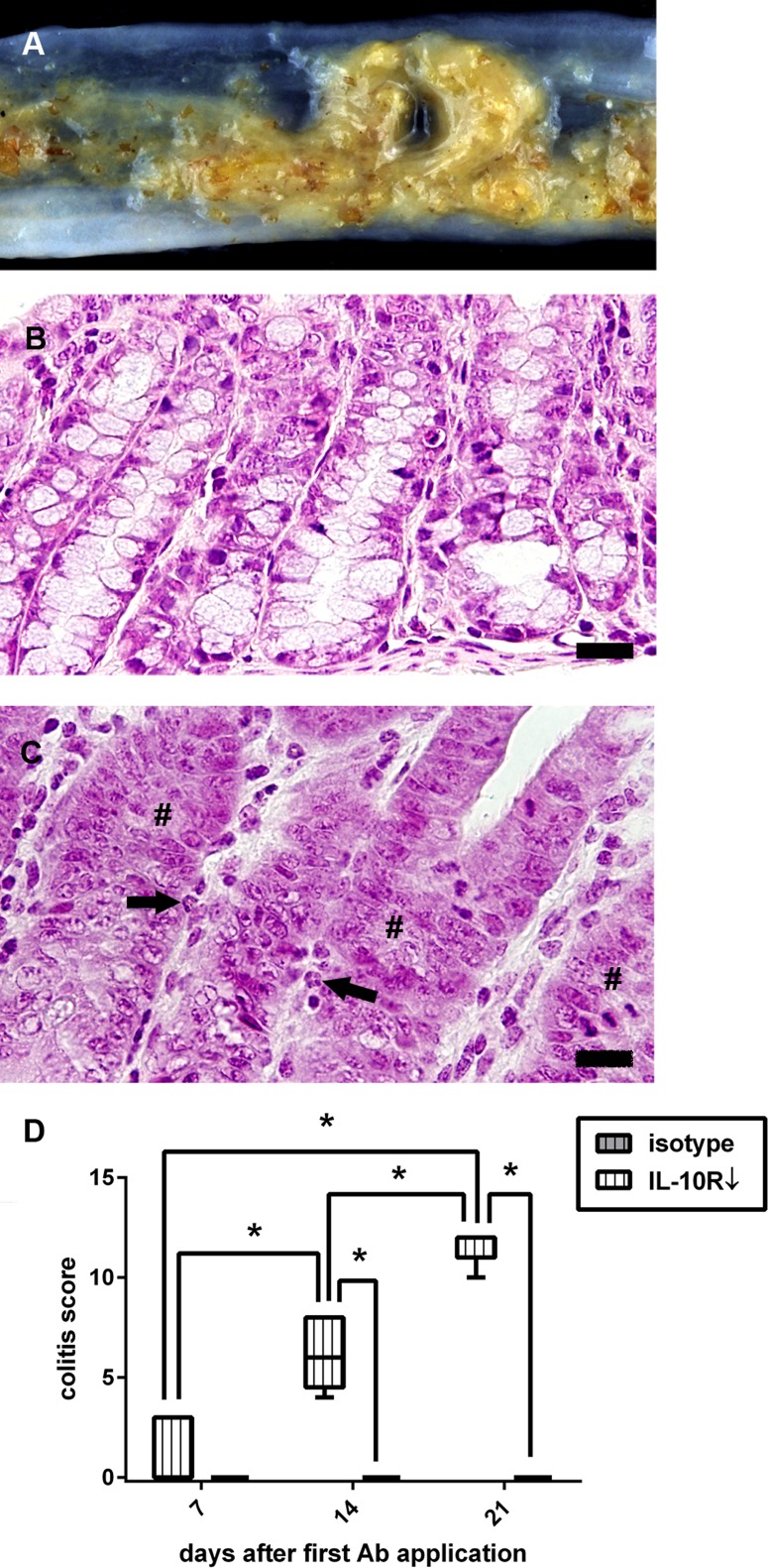
Fig 10. Enteric disease following interleukin-10 receptor (IL-10R) blockade in non-infected SJL mice.
(A) Unformed feces and mucosal edema in an animal at 14 days after onset of IL-10R antibody (Ab) treatment. (B) Colon of a control animal (group “isotype”) at day 21 with normal mucosal architecture. (C) Colon of an animal receiving IL-10R Ab (group “IL-10R↓”) at the same time point. Note infiltration of neutrophilic granulocytes (arrows) and loss of goblet cells (#). B, C: H&E staining, bar = 20 μm. (D) IL-10R blockade causes progressive colitis compared to control animals. grey box with vertical lines = control animals (group “isotype”), white box with vertical lines = IL-10R blocked animals (group “IL-10R↓”). Box and whisker plot display median, minimum and maximum values as well as upper and lower quartiles, 5 animals used at all investigated time points, Wilcoxon rank-sum tests, * = p < 0.05.

