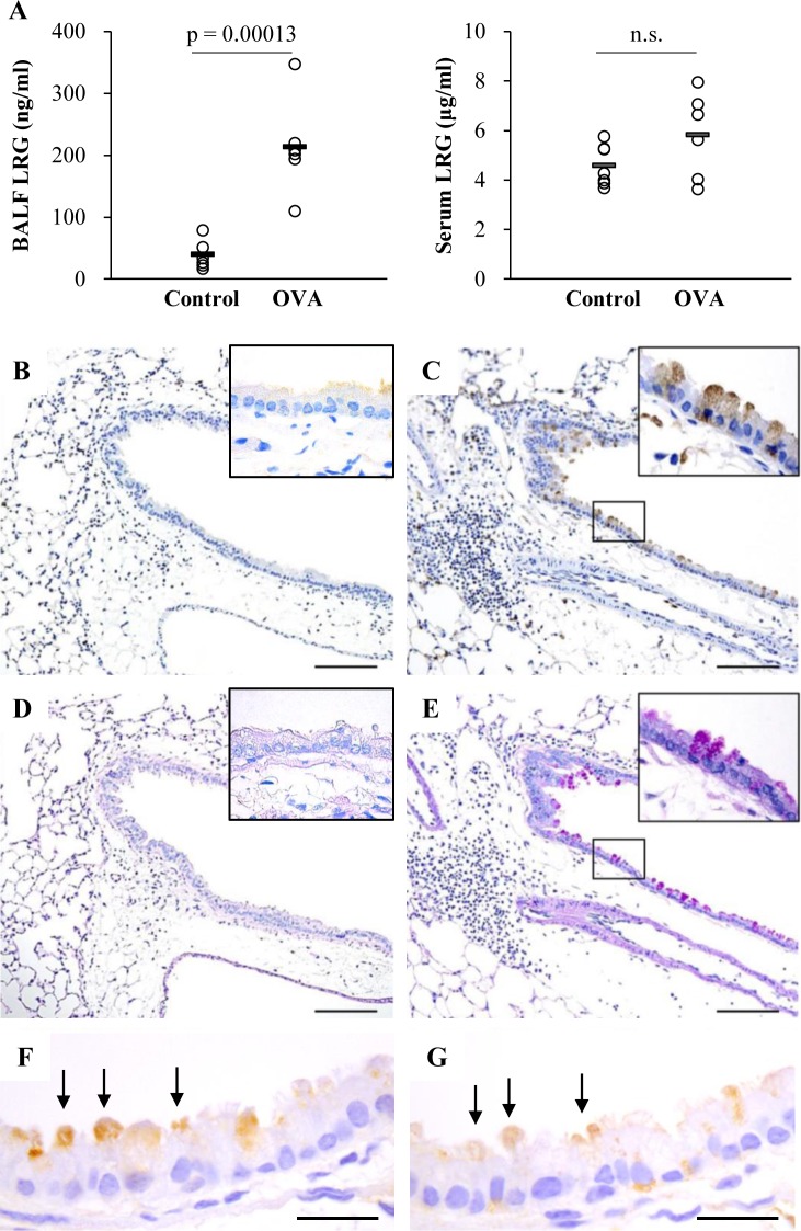
Fig 2. Detection of LRG in BALF, serum and lung section in a murine model of asthma.
A) Levels of LRG in BALF and serum in a mouse model of asthma. Concentrations of BALF and serum LRG were determined by ELISA. The Student’s t-test was used for statistical analysis. The individual values are provided in S3B–S3E File) Localization of LRG in mouse lung. Paraffin sections of the lung from control (B and D) and OVA-treated (C and E) mouse were stained with anti-LRG antibody (B and C) and PAS (D and E). Scale bar, 100 μm. F and G) Immunohistochemisry of MUC5AC (F) and LRG (G) of the lung from OVA-treated mouse. Arrows show MUC5AC (F) or LRG (G) positive cells. Scale bar, 20 μm

