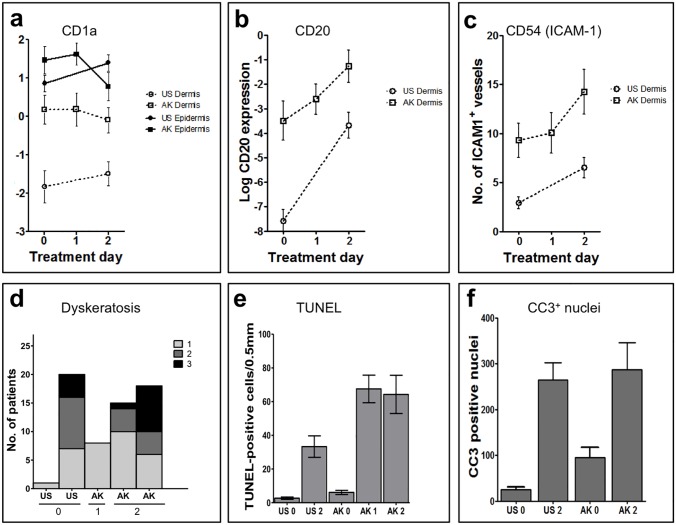
Fig 5. Topical ingenol mebutate 0.05% gel activates cutaneous blood vessels, attracts B-cells, and induces epidermal cell death.
Automated histomorphometric analyses on all samples from all patients (n = 26) were performed to quantitate expression of CD1a+ dendritic cells (A), CD20+ B-cells (B), and CD54 (ICAM-1) expressing cutaneous blood vessels and inflammatory cells within the dermis (C). Expression of both CD20 and CD54 increased rapidly and strongly upon treatment with ingenol mebutate, and actinic keratosis (AK) lesions showed generally higher expression levels compared with uninvolved skin. (D) Dyskeratotic (dead) keratinocytes within the epidermis of all samples (n = 26 patients; five biopsy specimens each) were assessed using a scoring system ranging from 0 (absent, not depicted), 1 (few dyskeratotic cells), 2 (several dead cells) to 3 (numerous necrotic cells). No necrosis was evident in the dermis. TdT-mediated dUTP-biotin nick end labeling (TUNEL)+ apoptotic cells (E) and cleaved caspase 3 (CC3)+ nuclei (F) were determined in biopsies from five patients. A full statistical overview is available in S1 Table.

