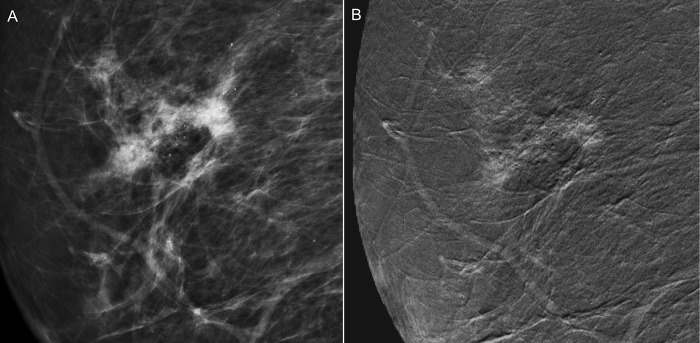
Fig 2. A 55-year-old woman receiving biennial mammographic screening with the finding of benign microcalcifications for 8 years was upgraded from BI-RADS category 3 to 4 in a recent examination because of increased microcalcifications.
(A) Low energy conventional mammogram on mediolateral oblique view showed segmental amorphous microcalcifications in the right breast; however the sonographic evaluation did not find any associated lesion. (B) CESM revealed a 3.3-cm irregularly shaped and outlined regional enhancement associated with the area of microcalcification. Subsequently, stereotactic core needle biopsy and surgery proved it to be an invasive ductal carcinoma.

